How is the flower pollen collected and processed?
The pollen is collected from two separate sites, the University of Delaware Botanical Gardens and apiary, and the Mount Cuba gardens and apiary. The flower pollen is collected by cutting the flower from the desired plant and storing it. When photographs of the pollen are needed, the dried and cut flower is put under a stereo microscope on a slide, and using a scalpel or scissors, the flower’s anthers are carefully opened. Ethanol is then dropped onto the exposed pollen to wash it out from the anther. Once a significant amount of the pollen is on the slide, the flower is stored for future use, and the pollen is allowed to dry. Once the ethanol is dried, the pollen is then dyed with a magenta stain in order to make the ornamentation and apertures stand out. A slide cover is then put over it, and the slide is labeled with the flower name, if it is from Mount Cuba or University of Delaware, and the date of slide preparation.
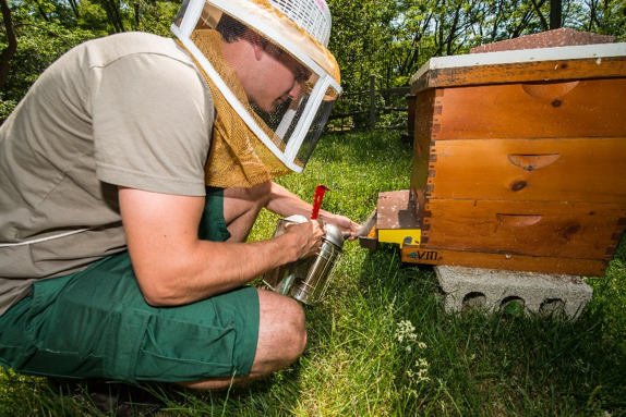
How is the bee pollen collected?
A pollen trap is placed on the hives both in the University of Delaware Apiary, and the Mount Cuba Apiary. The pollen trap is separated into two main components; the grid screen which knocks pollen off of the honey bees as they re-enter the hive from a day of foraging, and the basket where the pollen pellets are gathered.
 Once trapping of the pollen is finished, the grid screen is moved to the upright position so that the honey bees can come and go freely, without pollen being knocked off. The pollen is collected from the tray and put into airtight bags, which are labeled with the apiary from which it came, the hive name, and the date. The pollen is then transported to the lab for processing.
How is the bee-collected pollen collected and processed?
The collected pollen is put into bags according to the apiary and hive from which it came, then brought back to the lab for sorting and photographing. The pollen is sorted by color using Kirk’s A Colour Guide to Pollen Loads of the Honey Bee. Then the pollen, while still unprocessed, but sorted by color, is photographed, and then placed into microcentrifuge tubes, labeled by color and hive number as well as the date it was collected from the hive.
The pollen, once sorted by color and organized into respective microcentrifuge tubes, is then hydrated with water and placed in a centrifuge until the pollen is a liquid consistency. The pollen is put onto a slide using a pipette, and dyed with magenta stain. A cover glass is put over the pollen, and the slide is labeled with the date of extraction from the hive, the date of slide preparation, hive from which it came, and the color of the pollen.
How are photographs taken of the pollen?
The light microscopy photographs are taken using a Leica Microsystems DM750, with a Leica ICC50 HD camera. The flower pollen and bee-collected pollen slides are placed under the microscope and photographed at 40x and 100x magnification. Photographs are taken of all the diagnostic characteristics: the tectum, the exine and ornamentation, and the apertures. Photographs are also taken in both the polar and equatorial viewpoints if possible. The size, shape, and dispersal unit of the pollen are all noted. These characteristics are compared between the identified flower pollen and the bee-collected pollen in order to get an identification of the pollen the honey bees are carrying into the hive, and by extension, its nutritional value.
A more in depth overview of the Pollen Protocols can be found here: Pollen Protocols
